Animal Ophthalmology Clinic in FL
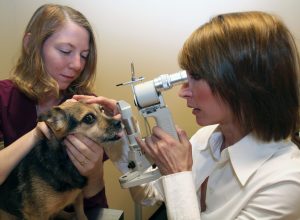
At Animal Eye Doctors, the well-being of pets is at the heart of every service provided. Specializing in animal ophthalmology, our clinic in Florida (FL) offers unparalleled expertise and state-of-the-art technology for all your pet’s eye care needs. Our team of highly trained ophthalmologists is dedicated to diagnosing and treating a wide range of eye conditions, ensuring that each pet receives the highest level of care.
Our Vet Ophthalmology Services for FL
Choosing a specialized animal ophthalmology clinic like ours over general veterinary services can make a significant difference in the treatment and management of your pet’s eye health. General veterinarians are skilled in a broad array of areas, but eye conditions often require more specialized knowledge and advanced techniques. At Animal Eye Doctors, our sole focus is on eye health, allowing our team to stay updated on the latest advancements in veterinary ophthalmology.
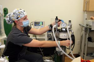
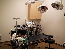
The clinic is equipped with cutting-edge technology designed to provide precise diagnoses and effective treatments. Advanced imaging systems, surgical equipment, and diagnostic tools enable our ophthalmologists to identify and address eye issues with exceptional accuracy. This investment in technology reflects our commitment to offering the best possible care for your pet.
Beyond technical expertise, compassionate care is a cornerstone of Animal Eye Doctors. Understanding that pets are family members, our team approaches each case with empathy and dedication. From the moment you and your pet walk through our doors, you will experience a warm, welcoming environment where your concerns are heard and addressed. We recognize that dealing with eye issues can be stressful, and our goal is to make the experience as smooth and reassuring as possible.
Choosing Animal Eye Doctors means placing your pet’s eye health in the hands of professionals who are passionate about their work. Our specialized focus, advanced technology, and compassionate care set us apart, making us the ideal choice for pet owners seeking the best for their furry companions.
To learn more or get the help that your pet needs, reach out to us today.
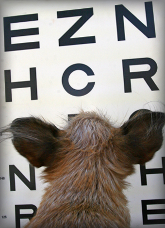
Animal Eye Doctors offers diagnosis and treatment of diseases and conditions relating to the eye and eyelids of your pet.
Diagnostic techniques and procedures to include:
- Slit lamp biomicroscopy
- Indirect ophthalmoscopy
- Tonometry/gonioscopy (glaucoma testing)
- Electroretinogram
- Ocular ultrasound
- Lid Mass removal and keratotomy for indolent ulcers performed with “local”
- Phacoemulsification (Cataract Surgery) with Lens Implants
- Eyelid surgery
- Grafting techniques for corneal disease
- Glaucoma Laser (Endolaser and Transcleral) and Drainage Implant Surgeries
- Eye Injuries
- Orthopedic Foundation for Animals (OFA) eye examinations/certification for inherited ophthalmic disease (formerly known as Canine Eye Registry Foundation/ CERF examinations)